
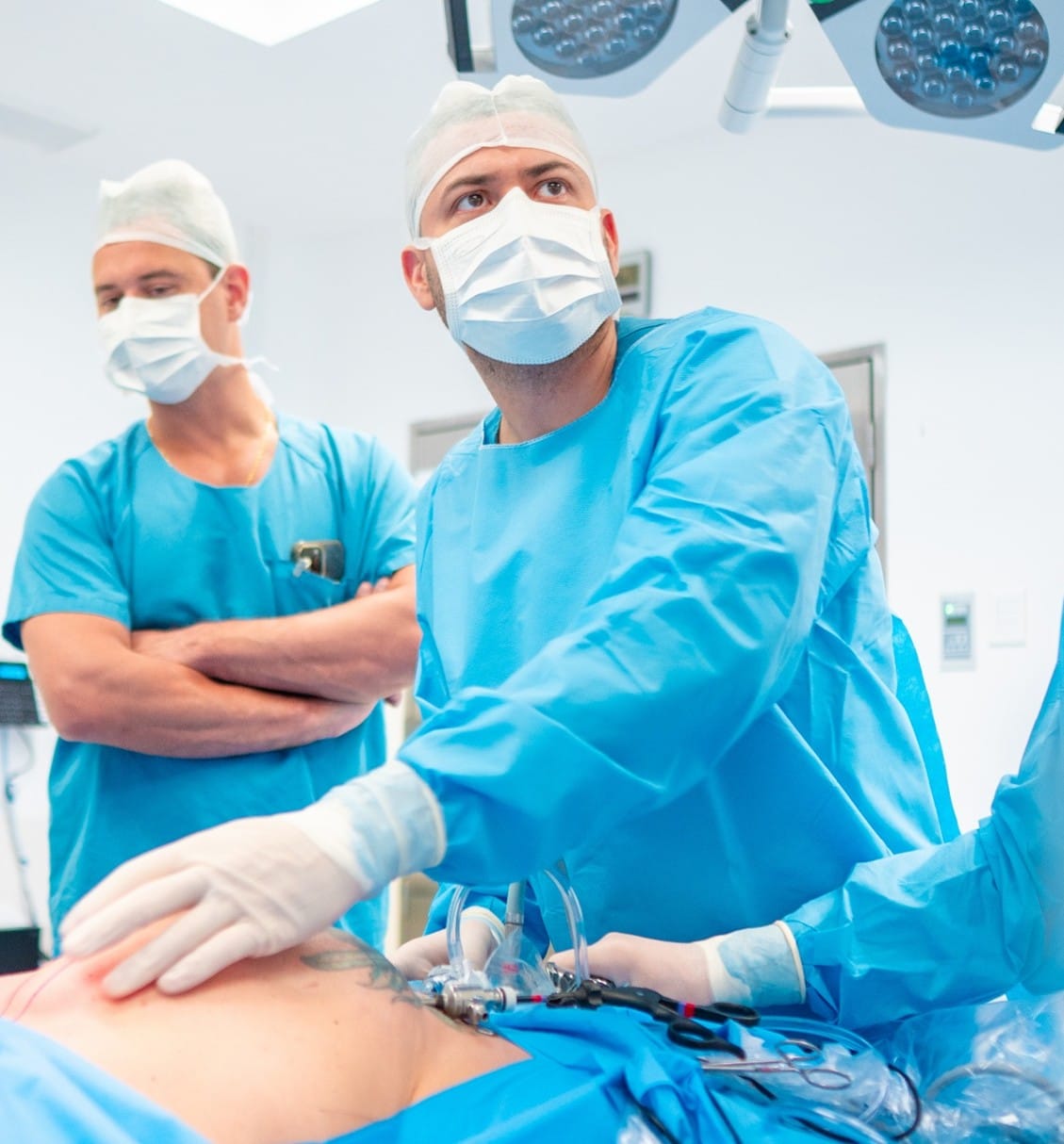
Rectus Diastasis Repair in Colombia – Surgical Core Restoration (Dr. José Agámez)
Pregnancy and major weight changes can stretch the abdominal wall and widen the midline — the “core” may lose its central support
Rectus diastasis is a structural condition, surgical indication depends on functional impairment, not appearance alone
Not all patients require surgery — decision-making is based on risk–benefit assessment
From a clinical standpoint, rectus diastasis is not just an aesthetic concern—it can be a functional problem of the abdominal wall. Surgical indication depends on symptoms, functional limitation, tissue quality, and a clear risk–benefit balance. Not all patients require surgery: mild diastasis with minimal impact may improve with targeted rehabilitation, while moderate-to-severe cases (or diastasis associated with hernias or significant laxity) may benefit from repair to restore core function and stability. I evaluate each case individually and explain the decision logic before recommending any procedure.
Concerned about whether your abdominal bulge may be a true hernia instead of rectus diastasis?
Learn how ventral and umbilical hernias are evaluated and treated
and understand the key differences before considering surgery.
What Is Rectus Diastasis? A Structural Separation of the Abdominal Wall
Rectus diastasis is defined as an abnormal widening of the linea alba, the connective tissue that joins the two rectus abdominis muscles at the midline. Rather than a true “hole” in the abdominal wall, it represents a loss of midline tension and mechanical integrity. From a clinical standpoint, this structural separation may alter force transmission across the core, affect posture, and reduce abdominal stability during movement or load-bearing activities.
Diagnosis is primarily clinical, based on physical examination while the patient activates the abdominal wall. In selected cases, ultrasound imaging is used to measure the inter-recti distance and evaluate tissue quality or associated hernias. Surgical indication depends not only on the measured distance but on functional symptoms, tissue laxity, coexistence of hernias, and a clear risk–benefit assessment.
How is rectus diastasis different from a ventral or umbilical hernia?
Although both conditions affect the midline, rectus diastasis does not involve a fascial defect through which abdominal contents protrude. In hernias, there is a true defect in the abdominal wall that may allow fat or bowel to protrude, sometimes requiring urgent repair. In rectus diastasis, the fascia remains continuous but stretched. However, both conditions can coexist, and identifying this distinction is essential when planning surgical repair.
When does rectus diastasis become clinically significant?
Not all diastasis requires intervention. Mild widening without symptoms may be managed conservatively with structured core rehabilitation. Clinical relevance increases when separation is associated with persistent abdominal bulging, lumbar discomfort, impaired force transmission, functional weakness, or recurrent midline hernias. Decision-making is therefore based on symptom severity, mechanical impairment, and patient goals rather than measurement alone.
When Is Rectus Diastasis Surgery Recommended? A Clinical Decision Framework
Surgical indication for rectus diastasis repair depends on functional impairment rather than appearance alone. From a clinical standpoint, the decision is based on midline mechanics, symptom severity, tissue quality, coexistence of hernias, and the likelihood of meaningful functional improvement after reconstruction.
Not all patients with visible abdominal separation require surgery. Many mild cases respond to structured rehabilitation. The objective is to intervene only when structural instability or progressive dysfunction justifies surgical correction.
Clinical Criteria Used in Decision-Making
1. Persistent functional instability
Surgery is considered when patients report ongoing midline bulging, core weakness, lumbar discomfort, or impaired force transmission that does not improve after appropriate rehabilitation.
2. Structural widening with poor tissue quality
When the linea alba demonstrates significant widening and reduced tensile resistance, conservative strengthening may not restore adequate midline tension.
3. Associated umbilical or midline hernia
The coexistence of a true hernia changes the mechanical environment of the abdominal wall. In these cases, combined repair may be indicated to prevent recurrence and restore stability.
4. Progressive worsening over time
Progressive separation, increasing instability, or worsening symptoms despite conservative management may justify surgical intervention.
5. When surgery is NOT recommended
Surgery may not be appropriate in cases of minimal separation without functional limitation, high surgical risk, unrealistic expectations, or when structured rehabilitation has not yet been attempted. Decision-making prioritizes safety, long-term durability, and proportional benefit.
Surgical Options for Rectus Diastasis: Technique Selection Based on Clinical Criteria
Midline Plication: Restoring Linea Alba Tension Without Cutting Muscle
What does plication actually correct?
Plication brings the rectus muscles back toward the midline by suturing the fascial layer (linea alba or anterior rectus sheath) to restore central tension. From a clinical standpoint, the target is not the muscle itself but the connective tissue that has widened and lost mechanical integrity. This step re-establishes load transfer across the abdominal wall and is foundational in most diastasis repairs.
When is plication alone sufficient—and when is it not?
Plication alone may be appropriate in selected patients with moderate separation, good tissue quality, and no associated hernia. However, it may be insufficient when the linea alba is markedly attenuated, separation is wide, tissue laxity is significant, or recurrence risk is elevated. Surgical indication depends on tissue behavior, functional impairment, and durability expectations—not on diagnosis alone.
Linea Alba Reinforcement (Selected Cases): Improving Long-Term Durability
When is reinforcement beyond sutures considered?
Reinforcement may be considered when the midline connective tissue is thin, lax, or under excessive tension—situations in which sutures alone may not provide durable repair. Examples include wide diastasis, poor fascial quality after major weight changes, recurrent midline defects, or coexistence of hernia. Matching the repair strategy to tissue quality is essential to reduce recurrence risk.
Does reinforcement always require mesh?
Not necessarily. Reinforcement may involve advanced suture strategies or, in carefully selected patients, mesh-based support to improve mechanical stability. Not all patients require mesh, and in some cases it is not indicated. The decision depends on anatomy, coexisting hernias, prior surgeries, infection risk, and the overall risk–benefit assessment.
Diastasis with Excess Skin or Fat: When Muscle Repair Alone Is Not Enough
Why can core repair be functionally successful but aesthetically incomplete?
Rectus diastasis repair restores midline stability and mechanical integrity. However, abdominal contour may remain suboptimal when significant skin laxity or localized adiposity is present. These are distinct problems: structural weakness versus soft-tissue envelope quality. When envelope quality limits the expected outcome, combined approaches may be considered to align functional correction with realistic contour goals.
When is a combined approach appropriate—and when should it be avoided?
Combination strategies may be appropriate when skin redundancy or adiposity would predictably limit contour improvement after muscular repair. However, not all patients benefit from combined procedures. If the primary objective is functional stabilization with minimal scarring—or if risk factors increase complication probability—a staged or limited approach may be safer. Clinical planning prioritizes safety, tissue perfusion, and individualized goals.
Diastasis with Umbilical or Midline Hernia: A Unified Abdominal Wall Strategy
Why repairing a hernia alone may not be sufficient
When a hernia coexists with rectus diastasis, the abdominal wall is mechanically weakened along the entire midline—not only at one focal defect. Repairing the hernia without restoring linea alba tension may leave abnormal force distribution unaddressed, increasing recurrence risk. Surgical planning therefore often involves addressing both problems within a single biomechanical strategy.
How is the surgical plan determined in combined cases?
The operative plan depends on hernia size and type, tissue quality, symptom profile, patient risk factors, and long-term durability considerations. Not all combined cases require the same approach: some benefit from minimally invasive repair, others from reinforcement, and some from staged procedures. Decision-making is individualized and based on restoring function while minimizing unnecessary complexity.
If your main concern is diastasis plus excess skin or localized fat, the surgical plan may include soft-tissue contouring in selected cases.
See the Spanish overview of MILA (minimally invasive lipoabdominoplasty)
for a simpler, patient-friendly explanation of when contouring is considered.
Minimally Invasive and Robotic Approaches: When Technology Adds Clinical Value
Rectus diastasis repair can be performed using minimally invasive techniques, including laparoscopic and robotic-assisted approaches. From a clinical standpoint, the choice of technology is not cosmetic—it is functional. Surgical planning depends on anatomical characteristics, tissue quality, coexistence of hernias, and the need for reinforcement. When appropriately indicated, minimally invasive techniques may reduce tissue disruption while maintaining mechanical restoration of the midline.
- Reduced soft-tissue trauma compared to open dissection in selected cases
- Improved visualization of the abdominal wall planes
- Precise midline reconstruction under magnified view
- Potential for lower postoperative discomfort
- Earlier return to light daily activity in many patients
- Smaller incisions and controlled tissue handling
- Enhanced anatomical accuracy during repair
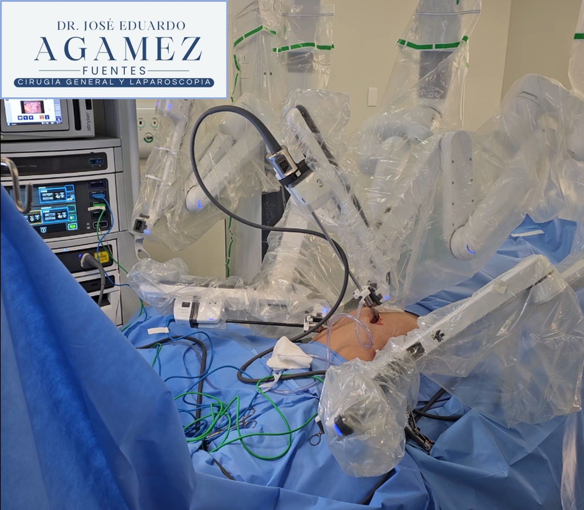
Robotic-Assisted Surgery (Da Vinci System): When Is It Indicated?
Robotic-assisted surgery provides enhanced three-dimensional visualization and articulated instrument control, which may improve precision in selected abdominal wall reconstructions. However, not all patients require robotic assistance. Its use depends on case complexity, prior surgeries, defect characteristics, and overall surgical objectives. In Colombia, robotic procedures are performed within accredited institutions where this technology is available, ensuring continuity of care and postoperative follow-up.
Recovery After Rectus Diastasis Repair: Functional Restoration and Clinical Oversight
Recovery after rectus diastasis repair is progressive and structured. From a clinical standpoint, the objective is not only wound healing but restoration of midline tension, force transmission, and core stability. Early mobility is encouraged while protecting the repair from excessive strain during the critical healing phase.
Not all patients recover at the same pace. Healing depends on tissue quality, surgical complexity, presence of reinforcement, associated hernia repair, and individual risk factors. Postoperative guidance is individualized to balance mobility, safety, and long-term durability of the repair.
Professional Perspective: How I Prioritize Safety and Functional Outcomes
1. Surgical indication is based on function, not appearance alone
Decision-making is based on symptom severity, mechanical impairment, and risk–benefit balance. Cosmetic improvement may occur, but functional restoration of the abdominal wall is the primary objective.
2. Not all patients require surgery
Mild diastasis without functional limitation may be managed conservatively. Surgery is considered when structural separation affects stability, quality of life, or is associated with hernia or progressive weakness.
3. Early movement is encouraged—but controlled
Patients are encouraged to walk early to reduce thrombotic risk and improve circulation. However, high intra-abdominal pressure activities are restricted during the initial healing period to protect the repair.
4. Core rehabilitation follows a structured progression
Rehabilitation progresses from breathing control and gentle activation to gradual strengthening. Sudden or aggressive core training too early may compromise repair integrity.
5. Reinforced repairs may require additional protection
When reinforcement or combined procedures are performed, postoperative restrictions may be slightly extended to optimize long-term durability.
6. Long-term stability depends on patient adherence
Weight stability, avoidance of abrupt strain, and proper reconditioning of the abdominal wall significantly reduce recurrence risk and improve functional outcomes.
7. The goal is durable midline restoration, not short-term results
A successful repair prioritizes mechanical integrity and long-term function over rapid cosmetic expectations. Surgical planning and recovery protocols are designed to ensure sustainable structural stability.
Frequently Asked Questions About Rectus Diastasis Repair in Colombia
Who is a candidate for rectus diastasis repair?
Candidates are typically patients with clinically significant separation that causes persistent midline bulging, functional weakness, lumbar discomfort, or impaired core stability—especially when conservative rehabilitation has not achieved adequate improvement. Surgical indication depends on symptoms, tissue quality, associated hernias, and risk–benefit assessment. Not all patients require surgery.
Can rectus diastasis be corrected with exercise alone?
Exercise can improve strength and motor control, but it does not always restore midline tension when the linea alba is structurally widened and tissue quality is poor. Mild cases may improve with structured rehabilitation, while moderate-to-severe cases (or those with hernias or significant laxity) may require surgery to restore mechanical integrity.
How is rectus diastasis diagnosed?
Diagnosis is primarily clinical, based on physical examination during abdominal wall activation and assessment of midline mechanics. In selected cases, ultrasound is used to measure inter-recti distance, evaluate tissue quality, and rule out associated umbilical or midline hernias that could change surgical planning.
What is the difference between rectus diastasis and a hernia?
Rectus diastasis is widening of the midline connective tissue without a true fascial defect. A hernia involves an actual defect through which fat or bowel can protrude and may carry risk of incarceration. The two conditions can coexist, and identifying both is important because a combined strategy may be required.
What surgical techniques are commonly used?
Most repairs involve midline plication to restore linea alba tension. In selected cases with poor tissue quality or high recurrence risk, reinforcement strategies may be considered. Technique selection depends on symptoms, tissue behavior, separation characteristics, coexistence of hernias, and surgical objectives.
Does minimally invasive or robotic surgery improve outcomes?
Minimally invasive approaches can reduce soft-tissue disruption in selected cases and allow precise midline reconstruction under magnified visualization. Robotic assistance may add value in complex reconstructions by improving instrument control and visualization. Not all patients require these technologies—use is determined by case complexity and risk–benefit balance.
Will the surgery leave visible scars?
Minimally invasive repairs typically use small incisions. Scar visibility depends on incision placement, skin type, and healing response. When combined procedures addressing skin redundancy are needed, scar patterns and trade-offs should be discussed explicitly to align the plan with realistic expectations and safety.
How long is recovery, and when can I return to exercise?
Recovery is progressive. Early walking is encouraged, while high-pressure core activities are restricted during early healing. Return to structured training is typically gradual and individualized based on repair complexity, reinforcement needs, and tissue quality. A staged core rehabilitation plan helps protect repair integrity and improve long-term durability.
Can rectus diastasis come back after surgery?
Recurrence is uncommon when technique selection matches tissue quality and postoperative guidance is followed. Risk may increase with major weight fluctuation, premature high-pressure activity, new pregnancy, or untreated associated hernias. Long-term durability depends on both surgical strategy and patient adherence during recovery.
When is surgery NOT recommended?
Not all patients require surgery. It may not be recommended when symptoms are minimal, functional limitation is low, conservative rehabilitation is likely to succeed, or surgical/anesthetic risk outweighs expected benefit. Decision-making should be transparent and based on clinical criteria rather than appearance alone.